


CORE FACILITY CELLULAR IMAGING
CEITEC Masaryk University
Kamenice 753/5
625 00 Brno CZ
 cellim_support(at)ceitec.muni.cz
 +420 54949 7085
+420 54949 6133
 @Ceitec_CellimCF
 ceitec_cellimcf
 Cellular Imaging Core Facility

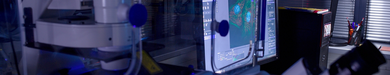
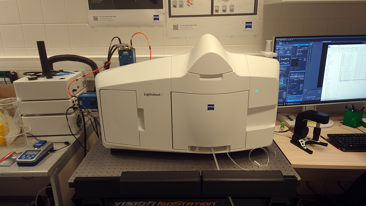
ZEISS Lightsheet 7
Description
The light sheet fluorescence microscopy (LSFM) has become a standard method to image large optically cleared specimens in toto and with subcellular resolution. It splits fluorescence excitation and detection into two separate light paths, with the axis of illumination perpendicular to the detection axis. That means you can illuminate a single thin section of the sample at one time, generating an inherent optical section by exciting only fluorescence from the in-focus plane. No pinhole is required. Light from the in-focus plane is collected as the pixels of a camera, rather than pixel by pixel as, for example, in confocal or other laser scanning microscopes. Parallelization of the image collection on a camera-based detector lets you collect images faster and with less excitation light than you would with many other microscope techniques. Zeiss Lightsheet 7 is equipped with dedicated optics, sample chambers and sample holders to accurately adjust to the refractive index of all common clearing methods and immersion media to allow imaging of large samples, even whole mouse brains.
Detailed description | |
Objectives | Detection optics: |
| Lasers | 405 nm, 488 nm, 561 nm, 638 nm |
Sample |
|
Filters for fluorescence | DAPI-GFP, GFP-Cy3, GFP-mCherry, GFP-Cy5, Cy3-Cy5 |
Camera | 2x PCO edge 4.2 sCMOS camera,1920 × 1920; |
Software | ZEN Black |